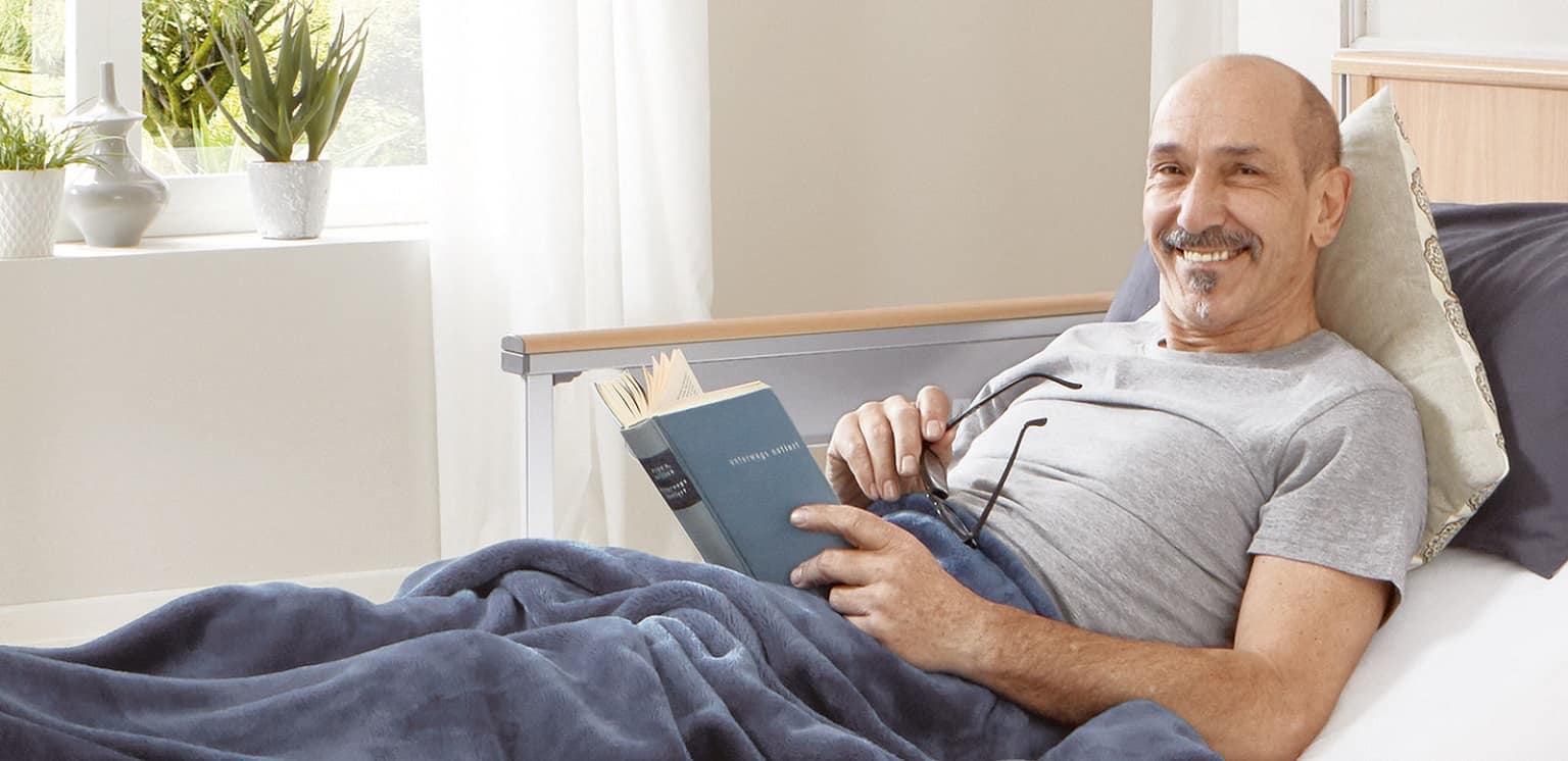
เครื่องช่วยฟังผู้สูงอายุ จำเป็นยังไง?
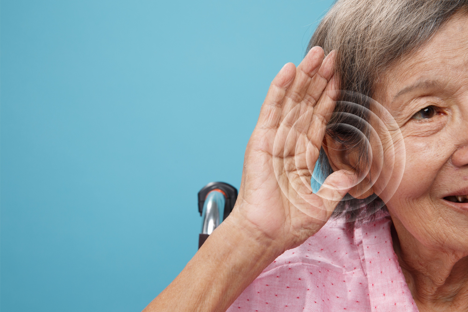
การได้ยินเสื่อมเป็นปัญหาสุขภาพที่พบได้บ่อยในผู้สูงอายุ โดยสถิติพบว่า ผู้สูงอายุมากกว่า 1 ใน 3 คนทั่วโลกมีปัญหาการได้ยินในระดับปานกลางถึงรุนแรง ซึ่งส่งผลกระทบต่อคุณภาพชีวิตในด้านต่าง ๆ มากมายเครื่องช่วยฟังเป็นอุปกรณ์ทางการแพทย์ที่ช่วยให้ผู้สูงอายุได้ยินเสียงได้ดีขึ้น โดยทำหน้าที่ขยายเสียงให้ดังขึ้นและปรับเสียงให้เหมาะสมกับระดับการได้ยินของผู้ใช้แต่ละราย เครื่องช่วยฟังผู้สูงอายุมีความจำเป็นในหลาย ๆ ด้าน ดังนี้ ช่วยให้ผู้สูงอายุสามารถสื่อสารได้อย่างมีประสิทธิภาพ : การสื่อสารเป็นทักษะที่จำเป็นต่อการดำรงชีวิตในทุก ๆ ด้าน ผู้สูงอายุที่มีปัญหาการได้ยินอาจประสบปัญหาในการได้ยินเสียงสนทนาของคนรอบข้าง ทำให้เข้าใจสิ่งที่คนอื่นพูดได้ยาก ซึ่งอาจนำไปสู่การเข้าใจผิด ความขัดแย้ง หรือความรู้สึกโดดเดี่ยว เครื่องช่วยฟังช่วยให้ผู้สูงอายุได้ยินเสียงสนทนาได้ดีขึ้น ทำให้สามารถสื่อสารได้อย่างมีประสิทธิภาพและเข้าใจสิ่งที่คนอื่นพูดได้อย่างถูกต้อง ช่วยให้ผู้สูงอายุสามารถมีส่วนร่วมในกิจกรรมต่าง ๆ ได้อย่างมีความสุข